


 An artist’s practice is often inspired and informed by what makes her head ache — be that a political stand, a gender issue or a medical concern. It is often an ache which is chronic and persistent. For the Dallas Art Fair’s featured artist, Paula Crown, the ache is actually in her head. Genetic migraines have pervaded her life and art. Hence, “Inside My Head: A Contemporary Self Portrait,” now at the Dallas Contemporary. Her video can be seen here.
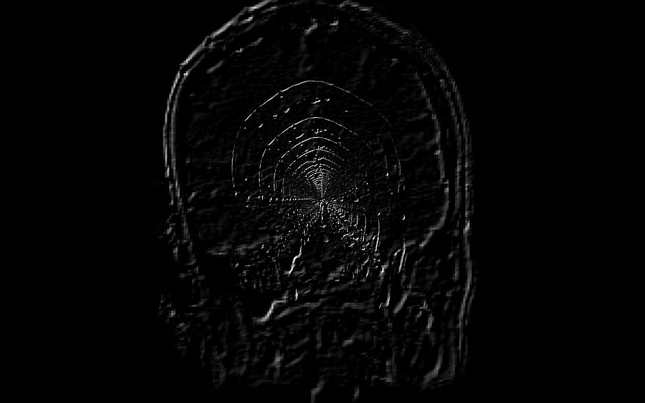
Beginning with the MRI scans of her brain, Crown uses digital technology to enlarge, embellish, enhance and manipulate those images. Using Photoshop and an open-source program processing, Crown can change perspective and more 2-D drawings into 3-D images.
Since “In My Head” combines science and art, I invited two physicians, Dr. Norma Melamed, a neurologist, and Dr. Jeffrey Glass, a psychiatrist-photographer, to walk through the installation with me. Although a few of the brain images are obvious, both doctors agreed that they would not have recognized several of the manipulated scans as brains. Most appeared as nebulous, organic shapes resembling planets or spiderwebs. Crown provides no titles to offer a hook for non-personal interpretations.
The artists says it is not important that we know the pictures are her brain — or any brain. “I view them as abstract forms and topologies that could be micro or macro in size.” Although Crown has said that landscapes capture her attention and she researches topologies and maps, there is not narrative or symbolism to her work. It’s the various shapes and patterns that interest her. My work is “just a connection between what is happening in our bodies and what is happening in the larger world,” she says. Interestingly, she has said that one picture might resemble a planet with waves. To Dr. Glass, the scan did look like a planet, but he also thought he saw the remains of truncated muscles and nerves from the original scan.
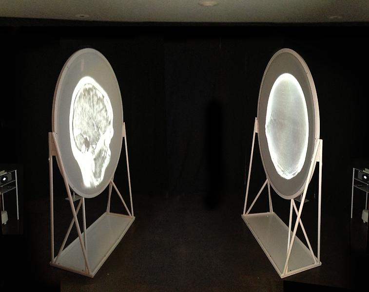
 In addition to the framed, manipulated scans, the Dallas Contemporary installation includes a room in which the viewer can be part of what Dr. Glass called an “inner-space trip,” allowing the visitor to experience something like an MRI. Visuals and audio envelop the viewer as he/she stands between two screens with varying projections of Crown’s brain rolling up and down and zooming in and out on the convex screens. The accompanying sounds (composed specifically for this piece) are not the hammering noise of an actual MRI scanner but a soothing violin, a musical interpretation of the activity of Crown’s brain. As the shapes change so does the music. “The music is my brain’s anatomy that was literally put into a software program and converted to sound,” Crown explains. “Then a violinist played to the animation of the brain scans.”
The soothing music resonated with Dr. Melamed — she uses sound to create a serene environment in her own office. She plays classical selections in the pink-painted room she reserves for patients requiring an EMG (electromyogram) — which involves shocks and needles.
Dr. Glass seemed to respond most to the video’s progression, which he said reminded him of swimming, “taking us on a journey through the layers of the brain, doctored with artistic license.” Each convex screen played different slices of the brain. The doctor equated the rolling pictures to those which the medical team would have seen as they scrolled down Crown’s images on their computers.
In this case, the doctors seemed to agree they saw no negative abnormalities or irregularities in the scans — just the positive abnormalities and irregularities of art.
COMMENTS